Question 1. Laboratory Practice: Serum Protein Electrophoresis - Question 1: (a) Define relative mobility (Rf); (b)…
Aminoàcids i estructura de les proteïnes
Context
Background: Electrophoresis separates biological macromolecules based on their net charge, shape, and size in an applied electrical field. At alkaline pH (such as in Tris-hippurate buffer, pH 8.8), serum proteins exist as polyanions because the buffer pH exceeds their respective isoelectric points (pI), causing them to migrate from the cathode towards the anode.
Objective: Separate serum protein fractions using cellulose acetate membrane electrophoresis, quantify electrophoretic mobility, evaluate technical parameters, and interpret pathophysiological protein patterns.
Question 1. Results calculation (relative mobility Rf and standards)
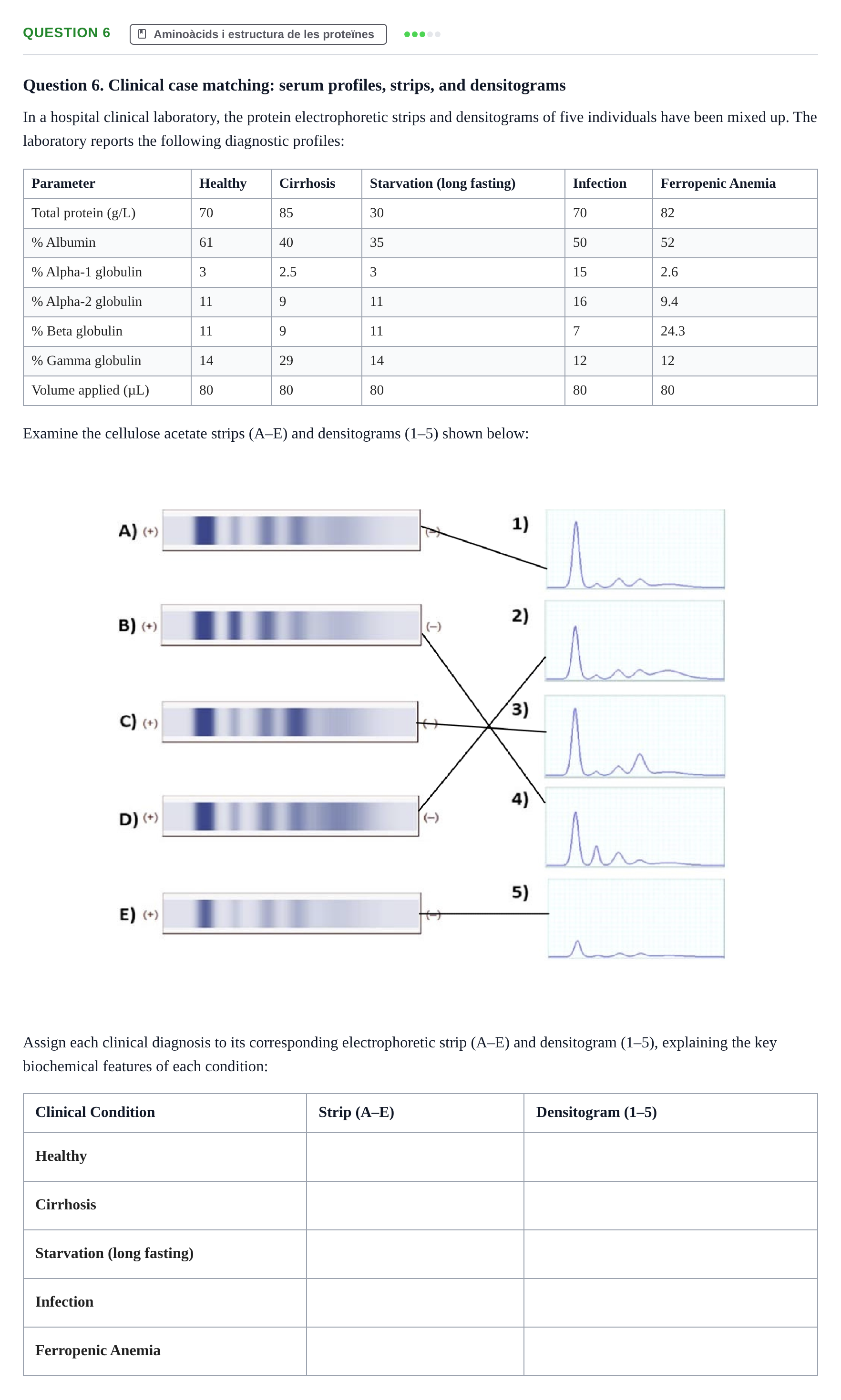
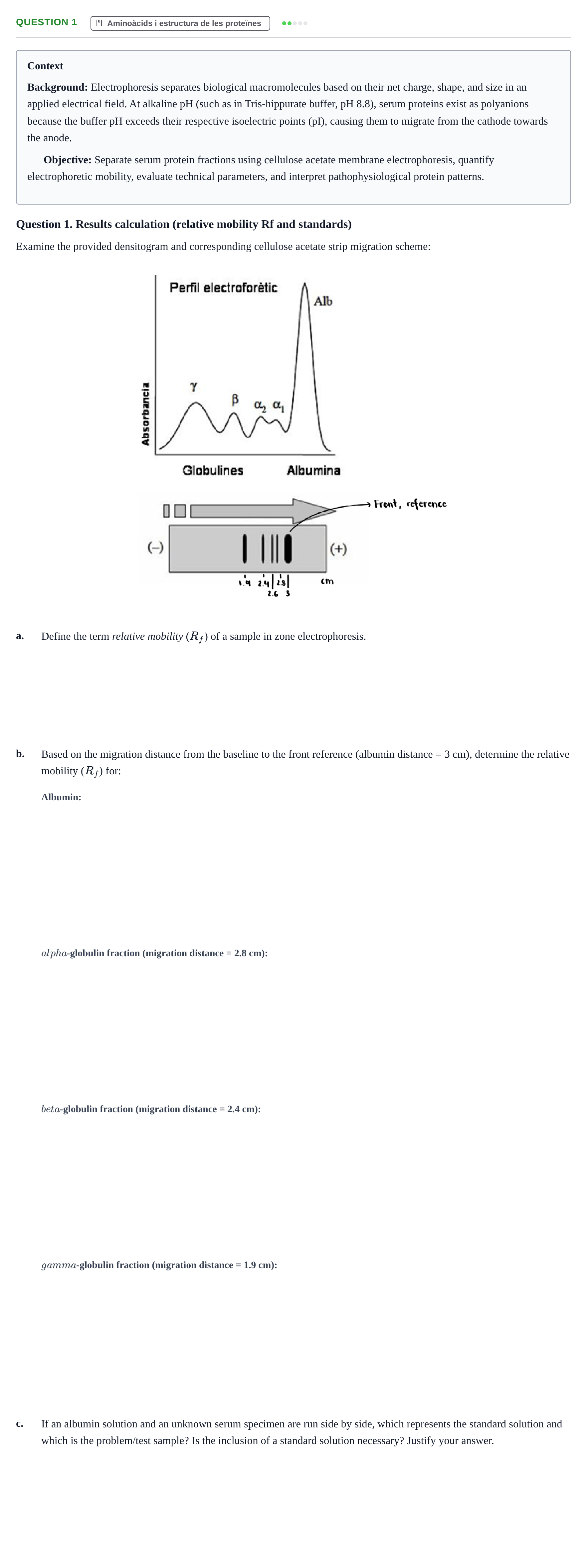
Examine the provided densitogram and corresponding cellulose acetate strip migration scheme:
a.
Define the term relative mobility () of a sample in zone electrophoresis.
b.
Based on the migration distance from the baseline to the front reference (albumin distance = 3 cm), determine the relative mobility () for:
c.
If an albumin solution and an unknown serum specimen are run side by side, which represents the standard solution and which is the problem/test sample? Is the inclusion of a standard solution necessary? Justify your answer.
Question 2. Explain serum protein behavior, net charge, and migration direction at buffer pH 8.8, pH 4.0, and pH 6.0.
Aminoàcids i estructura de les proteïnes
Question 2. Protein charge and migration at different pH levels
Explain the behavior and ionization state of serum proteins when placed in an electrophoresis buffer:
a.
What happens to serum proteins when immersed in the working electrophoresis buffer (pH 8.8)? Toward which electrode do they migrate?
b.
What charge would the proteins acquire and in which direction would they move if the buffer pH were set to pH 4.0?
c.
What migration behavior would be observed at pH 6.0?

Question 3. Predict the effect on the resulting densitogram and acetate strip if the volume of serum sample applied is…
Aminoàcids i estructura de les proteïnes
Question 3. Effects of increasing sample volume on electrophoresis
Predict the effect on the resulting densitogram and acetate strip if the volume of serum sample applied is increased:
a.
The separation and resolution between peaks will be:
b.
The intensity of the stained bands and the peak heights in the densitogram will be:
Question 4. Select the appropriate reagent or enzyme from the list (CNBr, trypsin, ninhydrin, DTT, urea, performic acid,…
Aminoàcids i estructura de les proteïnes
Question 4. Reagents used in protein biochemical analysis
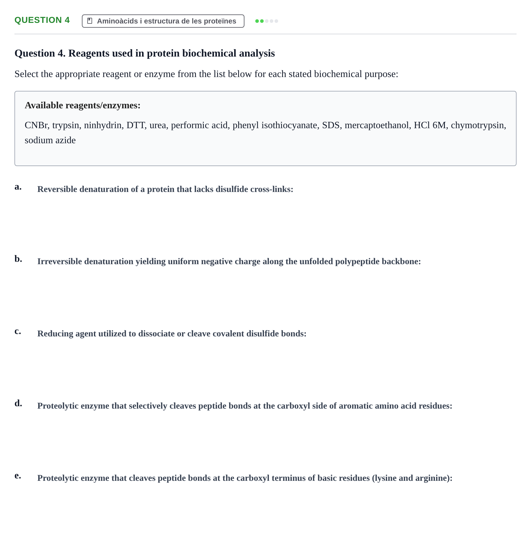
Select the appropriate reagent or enzyme from the list below for each stated biochemical purpose:
Available reagents/enzymes:
CNBr, trypsin, ninhydrin, DTT, urea, performic acid, phenyl isothiocyanate, SDS, mercaptoethanol, HCl 6M, chymotrypsin, sodium azide
a.
b.
c.
d.
e.
Question 5. Interpret a standard normal human serum densitogram to identify: (a) the protein fraction with the slowest…
Aminoàcids i estructura de les proteïnes
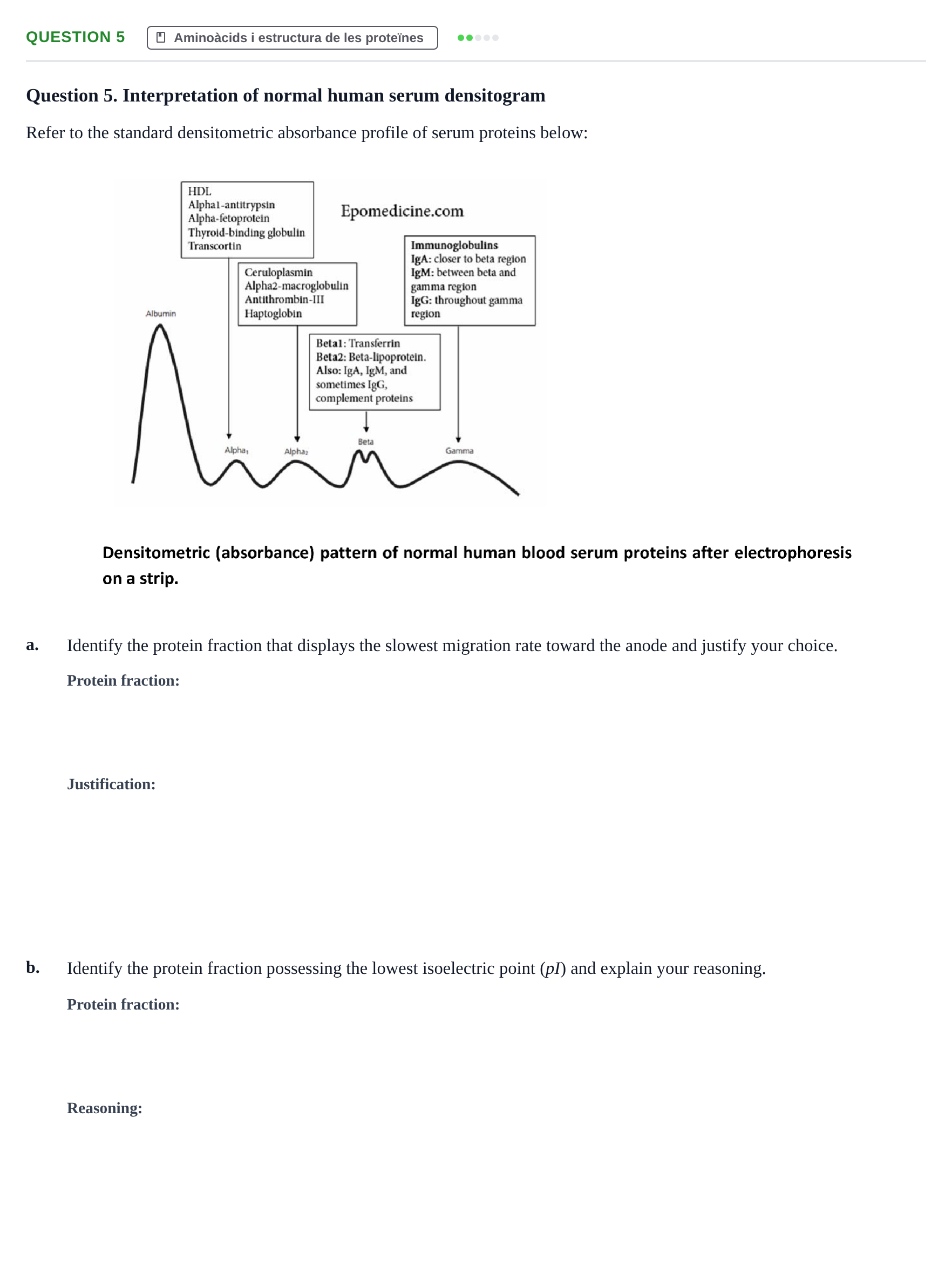
Question 5. Interpretation of normal human serum densitogram
Refer to the standard densitometric absorbance profile of serum proteins below:
a.
Identify the protein fraction that displays the slowest migration rate toward the anode and justify your choice.
b.
Identify the protein fraction possessing the lowest isoelectric point ( pI) and explain your reasoning.
Question 6. Assign each clinical diagnosis (Healthy, Cirrhosis, Starvation, Infection, Ferropenic Anemia) to its…
Aminoàcids i estructura de les proteïnes
Question 6. Clinical case matching: serum profiles, strips, and densitograms
In a hospital clinical laboratory, the protein electrophoretic strips and densitograms of five individuals have been mixed up. The laboratory reports the following diagnostic profiles:
Parameter · Healthy · Cirrhosis · Starvation (long fasting) · Infection · Ferropenic Anemia
Total protein (g/L) · 70 · 85 · 30 · 70 · 82
% Albumin · 61 · 40 · 35 · 50 · 52
% Alpha-1 globulin · 3 · 2.5 · 3 · 15 · 2.6
% Alpha-2 globulin · 11 · 9 · 11 · 16 · 9.4
% Beta globulin · 11 · 9 · 11 · 7 · 24.3
% Gamma globulin · 14 · 29 · 14 · 12 · 12
Volume applied (µL) · 80 · 80 · 80 · 80 · 80
Examine the cellulose acetate strips (A–E) and densitograms (1–5) shown below:
Assign each clinical diagnosis to its corresponding electrophoretic strip (A–E) and densitogram (1–5), explaining the key biochemical features of each condition:
Clinical Condition · Strip (A–E) · Densitogram (1–5)